Wasserthal, L.T. & Wasserthal, W. (1977b): Innervation of heart and alary muscles in Sphinx ligustri L. (Lepidoptera). A scanning and transmission electron microscopic study. - Cell Tissue Res. 184: 467-486.
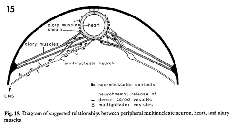
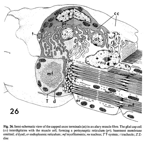
The origin and orientation of the heart nerves in Sphinx ligustri and Ephestia kuehniella were investigated by scanning electron microscopy using a special technique which involved pinning the dissected specimens on a stabilizing metal pad. The heart and alary muscles in Sphinx particularly their caudal extremity were also examined by transmission electron microscopy. The alary muscles form an incomplete sheath around the heart with a mainly longitudinal fibre orientation, e.i. antagonistically to the fibres of the heart itself. The heart and alary muscles are multiterminally innervated by branches of the transverse segmental nerves. All branches contain a single electron lucent axon; the thickest branches also possess several neurosecretory axons. Swellings of the segmental nerves may indicate the position of nerve cell bodies. There are no lateral heart nerves. Only one type of neuromuscular junction is abundant in the alary muscles but less frequently found in the heart. The terminals originate from the central axon only. They are capped by glial cells, which interdigitate with the muscle cells. They penetrate into the T-system toward the Z-discs and form a complex intercellular space system. Exocytosis of dense-cored vesicles into this "perisynaptic reticulum" seems likely. Sites of neurohaemal release are distributed along the nerve branches and special nerve endings occur at the level of the ostia. The possible nervous influence upon heart activity is discussed.